
FIG 1.
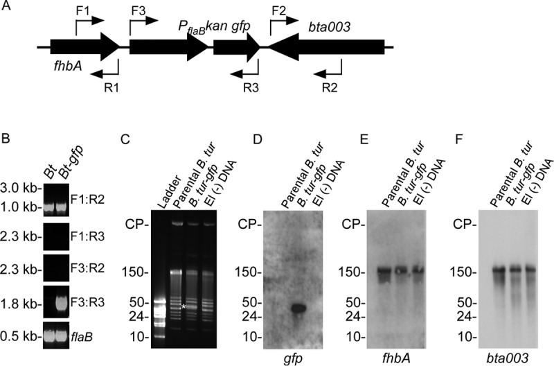
Integration of PflaB kan-gfp into B. turicatae. (A) PflaB kan-gfp was initially targeted for insertion between fhbA and bta003 with primer locations shown as thin arrows. (B) PCR was performed using wild-type B. turicatae (Bt) or B. turicatae-gfp (Bt-gfp) with primer combinations shown on the right of each gel image. (C to F) Pulsed-field electrophoresis (C) and Southern blotting (D to F) were performed to determine the recombination location of PflaB kan-gfp within the B. turicatae genome. (C) A shift in molecular weight was observed for a 40-kb linear plasmid of B. turicatae-gfp (white asterisk), which was not observed in the parental strain (parental B. tur) or B. turicatae that was electroporated without DNA (−DNA). (D to F) A probe designed for gfp localized the gene to a 40-kb linear plasmid (D), while fhbA and bta003 remained localized to lp150 (E and F). The probes for gfp, fhbA, and bta003 are shown beneath each Southern blot image. Molecular weights and circular plasmids (CP) are shown on the left of the gels and Southern blot.