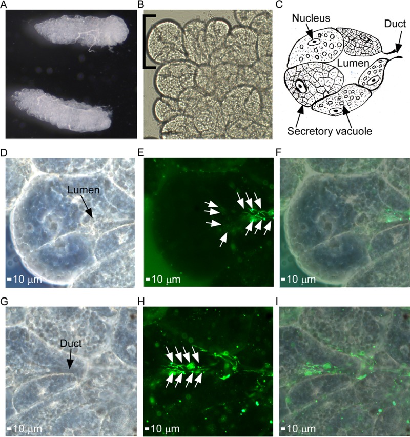
FIG 5.

Demonstration of salivary gland colonization by B. turicatae-gfp. (A) Intact salivary glands were excised from infected O. turicata ticks. (B and C) Agranular acini along the peripheral margin of the posterior region of the salivary gland (B) were assessed for spirochete colonization based upon location of the lumen and efferent duct, as illustrated in the artistic rendition of salivary gland tissue (C). Each acinus is composed of secretory cells that produce saliva, which drains into the lumen and through the efferent duct. A single acinus is shown in panels D to I and was viewed with a 63× oil immersion objective. (D to F) Spirochete colonization in the acini lumen is demonstrated. A dark-field image shows the acinus structural outline and location of the lumen (D), followed by fluorescent-filtered image showing localization of fluorescing spirochetes (E), indicated by white arrows, and an overlay of dark-field and fluorescent images (F). (G to I) Spirochete colonization in the efferent duct region in the same acinus is also demonstrated. Shown is a dark-field image of the acinus structural outline and location of the efferent duct (G), followed by fluorescent-filter image showing the localization of fluorescing spirochetes (H), as indicated by the white arrows. (I) An overlay of dark-field and fluorescent images is also shown.
