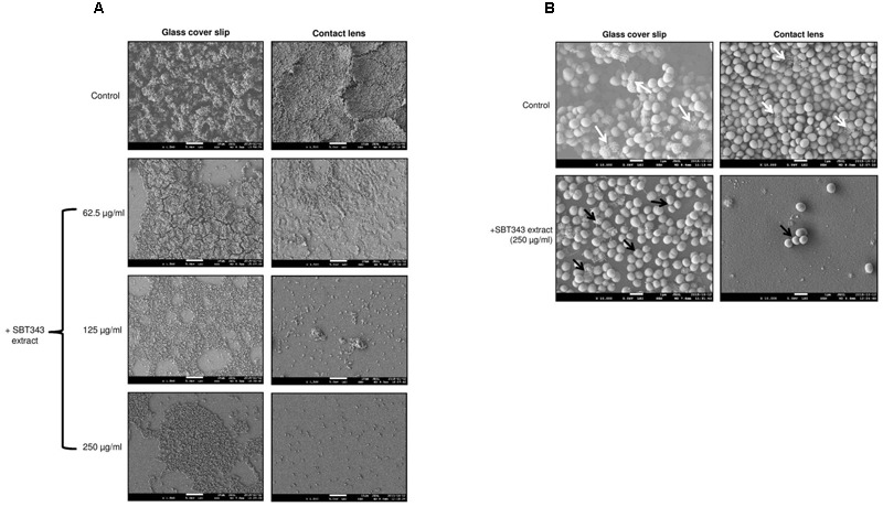
FIGURE 2.

Representative SEM images of staphylococcal biofilm on (A) Glass cover slip and contact lens at X1500 magnification. (B) Glass cover slip and contact lens at X10000 magnification. The white arrows indicate the presence of fibrous, net like structures which are typical features of PIA-dependent biofilms and the black arrows indicate the absence of the same. S. epidermidis RP62A treated with the extract from modified ISP2 medium (250 μg/ml) served as the appropriate control in the microscopy experiments.
