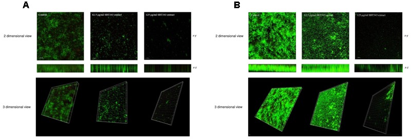
FIGURE 3.

Confocal laser scanning microscopy analyses of staphylococcal biofilm in the presence of the SBT343 extract, visualized by fluorescence vital dye. Images were acquired by Leica TCS SP5 with a 20X objective lens (scale bar 30 μm). (A) Biofilm images on the glass cover slip. (B) Biofilm images on contact lens. In (A,B), compressed z series (top), where multiple x–y planes from top to bottom of the biofilm are combined and the smaller image (bottom) represents the compressed x–z (side) of the biofilm. The thickness of the biofilms in control was 28.7 μm, whereas after extract treatment it was only 15.1 μm (62.5 μg/ml) and 4.53 μm (125 μg/ml) on the glass cover slips. Similarly, the thickness of the biofilms in the extract treated contact lens samples was only 13.6 μm (62.5 μg/ml) and 7.55 μm (125 μg/ml).
