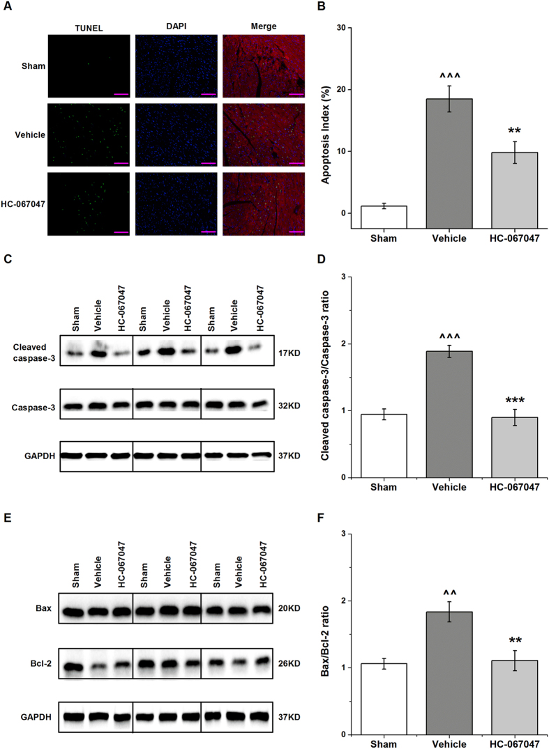
Figure 5. TRPV4 antagonist HC-067047 reduced cardiomyocyte apoptosis in a mouse model of myocardial I/R.
The TRPV4 antagonist HC-067047 (10 mg/Kg) was intraperitoneally injected at 1 h after reperfusion. (A) Representative photographs of TUNEL-stained heart sections from different groups at 4 h after reperfusion. Apoptotic nuclei were identified by TUNEL staining (green), cardiomyocyte by anti-sarcomeric actin antibody (red), and total nuclei by DAPI staining (blue). Scale bar: 100 μm (B). Percentages of TUNEL-positive nuclei over total number of nuclei. n = 8/group, ^^^P < 0.001 vs sham, **P < 0.01 vs vehicle. (C) Representative photographs of cleaved caspase-3 in groups by western blot after 4 h reperfusion. Full-length blots/gels are presented in Supplementary Fig. 6. (D) Cleaved caspase-3 in myocardium was assessed and the values were normalized to sham, n = 6/group, ^^^P < 0.001 vs sham, ***P < 0.001 vs vehicle. (E) Representative photographs of Bcl-2 and Bax in groups after 4 h reperfusion. Full-length blots/gels are presented in Supplementary Fig. 7. (F) The results were expressed as ratio of Bax/Bcl-2. n = 6/group, ^^P < 0.01 vs sham, **P < 0.01 vs vehicle.

