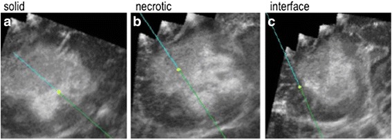
Fig. 2.

An example of the digital ultrasound images obtained from a WHO grade IV lesion. Biopsies were taken from the solid (a) and nectrotic (b) regions and the infiltrating interface (c) of a WHO grade IV lesion. On these ultrasound images, the blue line represents the biopsy needle, whilst the yellow circle is the tip, with a projected tip extension shown beyond the tip/yellow circle
