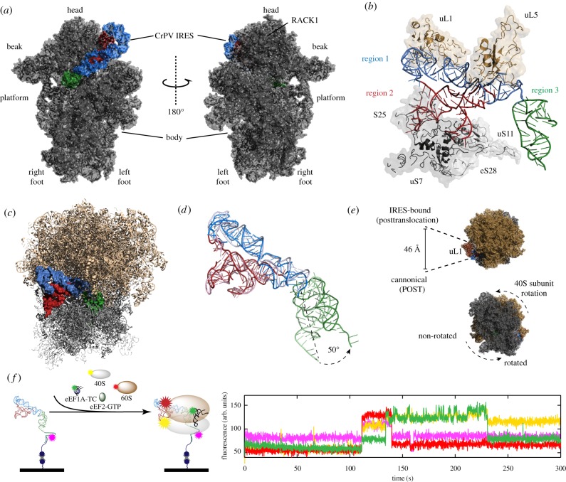
Figure 3.
Structure and dynamics of the ribosome-bound CrPV IRES. (a) 40S : IRES structure [46] (PDB entry 4V92). The 40S subunit inter-subunit face (left) and solvent face (right) structures are annotated with features of the small subunit. The IRES is coloured by region: region 1 (blue); region 2 (red); region 3 (green). (b) Structure of the 40S-bound CrPV IRES and IRES-interacting 40S (grey) and 60S (brown) ribosomal proteins [46] (PBD entry 4V92). (c) Structure of the 80S : IRES complex [46] (PBD entry 4V92). (d) Alignment of the pre- [28] (PBD entry 2NOQ) and post- [49] (PBD entry 4D5N) pseudotranslocated IRES. The post-pseudotranslocated IRES (dark colours) adopts a more extended conformation compared with the pre-pseudotranslocated IRES (light colours). (e) 60S subunit solvent face view of the 80S : IRES complex structure (top) highlighting L1 stalk displacement of the IRES-bound, post-pseudotranslocated state [49]. The 40S subunit solvent face view of the 80S : IRES complex structure showing 40S subunit rotation. (f) Observation of CrPV IRES-mediated initiation using single-molecule fluorescence. A schematic representation (left) of a ZMW single-molecule fluorescence delivery experiment where fluorescently labelled 40S (yellow) and 60S (red) subunits, eEF1A-TC tRNAPhe-Phe (green) and eEF2-GTP are co-delivered to surface-immobilized CrPV IRES-Cy5.5 (magenta). In the example trace from the experiment (right), simultaneous arrival of the 40S and 60S subunits (burst of red and yellow fluorescence at 110 s) to the immobilized IRES is followed by tRNA binding to the 80S : CrPV IRES complex (burst of green fluorescence at 130 s). Panel (f) has been reprinted with permission from Petrov et al. [29].

