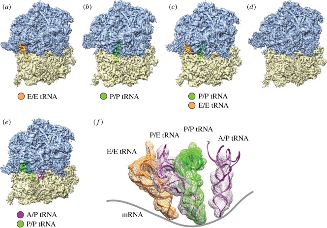
Figure 2.
Cryo-EM reconstructions of the P. falciparum 80S ribosomes purified from cell extract [93]. (a–d) Density maps of the P. falciparum 80S ribosome in non-rotated states. (a) Bound with E-tRNA (approx. 97 000 particles) at an average resolution of 4.7 Å; (b) bound with P-tRNA (approx. 14 500 particles) at 6.7 Å; (c) bound with P/P- and E/E-tRNAs (approx. 14 500 particles) at 6.7 Å and (d) without tRNAs (approx. 32 000 particles) at 5.1 Å. (e) Rotated state (approx. 23 000 particles) at 5.8 Å resolution; 60S subunits are coloured blue and 40S subunits are yellow. (f) Positions of tRNAs for all 80S complexes in (a–d). Structures and positions of E-, P- and P/E-tRNA were obtained by MDFF fitting, and the structure and position of A/P-tRNA was taken from the existing model, PDB 3J0Z, rigid-body fitted into the segmented map in UCSF Chimera. Contours of cryo-EM densities are displayed in mesh; structures of tRNA are displayed as ribbons and the mRNA path has been added as a cartoon. (Reproduced with permission from [93].)

