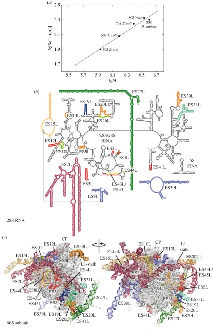
Figure 2.
Dependence of the sedimentation coefficient of the ribosome particle on molecular weight (a). Graphic kindly provided by S. Agalarov. Secondary structure of the human 28S RNA with coloured expansion segments (b). Molecular models of the 60S subunits from H. sapiens with ES coloured as in (b). Landmarks include the central protuberance (CP), L1-stalk and P-stalk. Figure kindly provided by D. Wilson.

