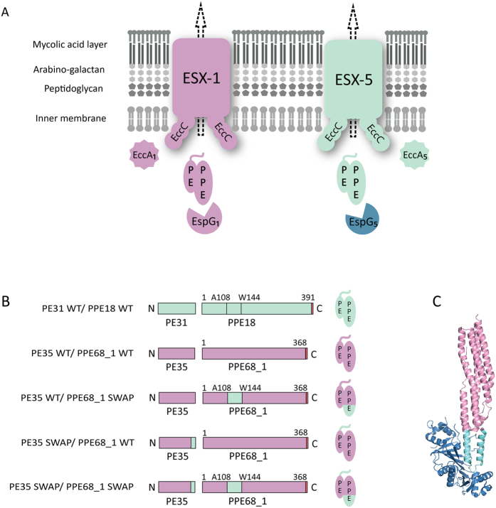
Figure 2. Model for substrate targeting and translocation in type VII secretion and a schematic representation of the different constructs used in this study.
(A) The current model for substrate targeting in type VII secretion. The mycobacterial cell envelope is composed of an inner membrane and an outer mycolic acid-containing membrane. EspG chaperones specifically recognize their cognate PE/PPE substrates. The general YxxxD/E secretion signal at the C-terminus of PE proteins is exposed for interaction with the secretion machineries. EccC is one of T7S membrane components containing three ATP binding domains and is probably involved in substrate recognition by the systems. (B) Schematic representation of the WT M. marinum ESX-5 substrates PE31/PPE18 WT (in cyan), the WT M. marinum ESX-1 substrates PE35/PPE68_1 (in pink) and derivatives used in this research. The colors indicate the origin of the different domains and the HA-tag is shown in red. (C) A representation of the structure of EspG5 (in blue) bound to the ESX-5 substrate PE25/PPE4111. The corresponding EspG binding region that is replaced in this study is indicated in cyan, while the remained parts of the PE/PPE substrates are highlighted in pink.

