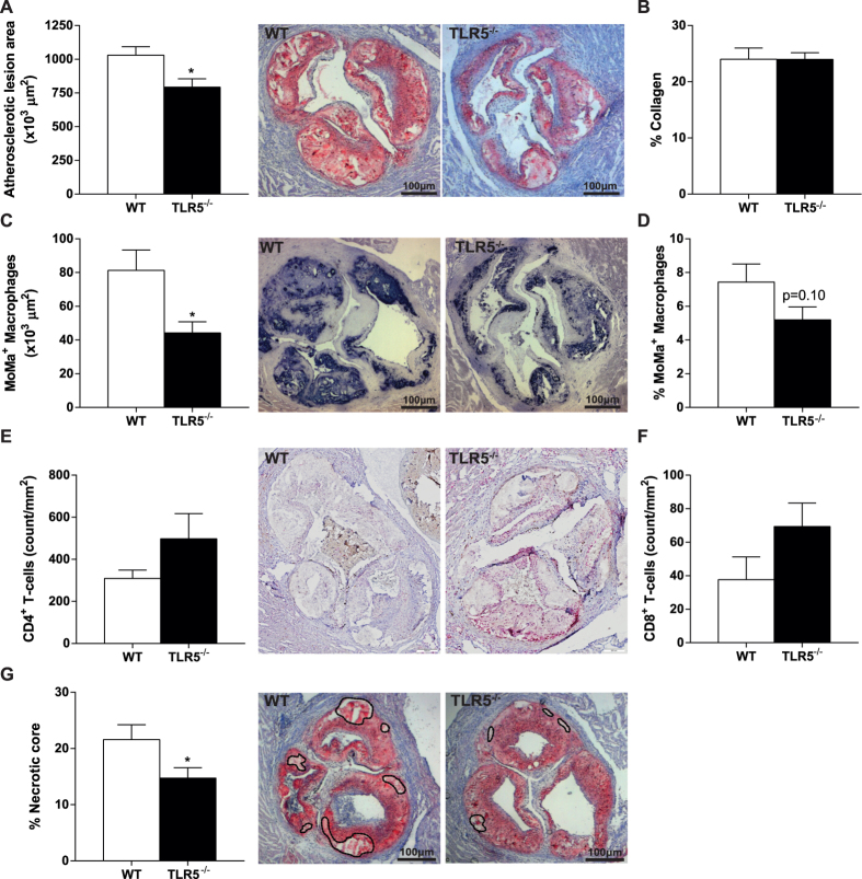
Figure 3. Atherosclerotic plaque size and composition.
After 12 weeks of high-fat diet, the atherosclerotic plaque area was significantly larger in WT mice compared to TLR5−/− mice (A). No difference between both groups was observed with respect to collagen content (B), yet the plaque of TLR5−/− mice showed a decrease in macrophage influx (C,D). Although not significant, TLR5−/− mice showed higher numbers of CD4+ (E) and CD8+ T-cells (F). The necrotic core expressed as a percentage of the plaque was significantly higher in WT mice (G); n = 12–13 per group (5-8 for CD4-8 staining). WT: wildtype, TLR5−/−: toll-like receptor 5 knockout, *p < 0.05.

