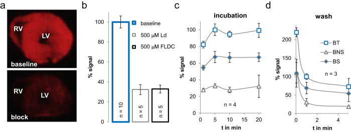
Figure 1. Ex vivo imaging using radiocaine autoradiography.
(a) Images of rat myocardial tissue slices (20 μm) incubated with radiocaine (top) or radiocaine and 500 μM fluorolidocaine (bottom). Both the left and right ventricle (LV & RV) are visible and surrounded by the myocardium, which displays a strong radioactive signal. Suppression of this signal by coincubation of fluorolidocaine demonstrates displaceable binding. (b) Comparison of the known SCN5A blocker lidocaine (Ld) and fluorolidocaine (FLDC) as competition ligands at the same concentration. Both ligands lead to the same level of non-displaceable binding. (c) Incubation times of 1, 5, 10 and 20 min were tested to investigate equilibrium conditions. After 5 min of radiocaine incubation, specific binding (BS) reached a constant level (BS = total binding BT − nonspecific binding BNS). (d) Washing times of 3 secs, 1 min and 5 min were tested to optimize signal to background conditions and investigate dissociation time course. Wash times of only 1 min are sufficient to reduce non-specific binding to a constant level. (error bars are presented as ± one standard deviation (stdev)).

