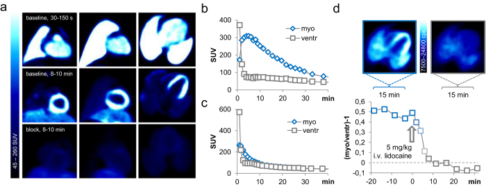
Figure 3. Radiocaine PET-imaging in non-human primates.
(a) Thoracic PET-images of a baboon injected with 5.08 mCi radiocaine. Upper panel shows summed images (30–150 seconds) from coronal, sagittal and transverse view with blood-filled heart and lungs. The middle panel shows the same scan summed from 8–10 min with a clear myocardial signal. The lower panel shows the same animal in a radiocaine scan treated with 5 mg/kg i.v. lidocaine 5 minutes prior to tracer injection. (b) TACs of the myocardium and ventricle of the baboon shown in the upper panel of (a). The larger organ (compared to rat) allowed using the ventricle as an internal reference for the radiocaine blood signal. (c) Analogous TACs of the baboon shown in the lower panel of (a). The competition ligand lidocaine, injected 5 minutes prior to radiocaine suppresses the myocardial signal to background levels. (d) An additional baboon investigated with a bolus + infusion paradigm and an in-scan lidocaine challenge. A dose of 5.0 mg/kg lidocaine was injected after the myocardial signal had reached a plateau. 10 min after this drug challenge, the myocardial signal was reduced to background levels.

