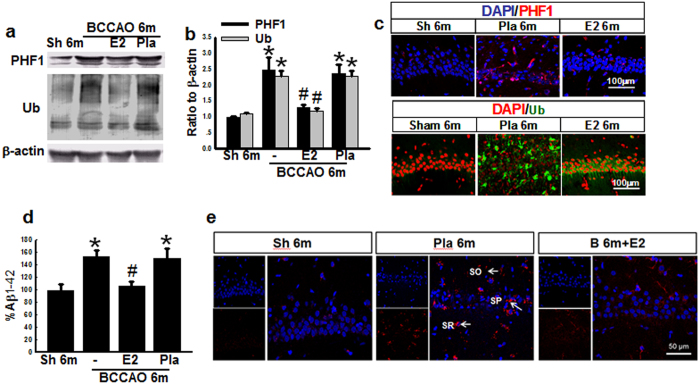
Figure 7. E2-treatment for 3 m decreases the protein expression of dementia biomarkers in hippocampal CA1 region 6 m after BCCAO.
Hippocampal samples from sham (Sh) 6 m, BCCAO 6 m, Placebo (Pla) 6 m and Estradiol (E2)-treated groups for 3 m were subjected to Western blot analysis for PHF1 and total Ub (a,b). (c) Representative photographs of immunofluorescent staining for PHF1 (Red) or total Ub (Green), with the nuclear stain, DAPI. (d) ELISA analysis for Aβ1-42, and (e) Immunofluorescent staining for Aβ1-42 (Red) and DAPI (Blue), indicating that Aβ1-42 is prominently deposited in hippocampal stratum radiatum (SR), stratum pyramidale (SP) and stratum oriens (SO) cell layers, and markedly decreased by E2 treatment. *P < 0.05 vs. sham 6 m group, #P < 0.05 vs Pla 6 m group. n = 5-6. Scale bar: 50 μm and magnification: 40×.

