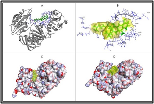
Figure 2.

Molecular interactions of murE and three of the prioritized drugs. Figure A displays the target protein in solid ribbon form, gray in colour with interacting amino acids highlighted in line form, blue in colour while the docked lymecycline is displayed in stick form, green in colour. The intermolecular hydrogen bonds are displayed in dotted lines, red in colour. The magnified view of the interactions for better clarity is provided in the Figure B; semi-transparent surface over lymecycline (yellow in colour) and interacting amino acids labelled with three letter code. The effective fitting of the acarbose and desmopressin in the active site groove of the murE is clearly displayed in figure C & D respectively; acarbose and desmopressin is highlighted in semi-transparent surface (yellow in colour) and displayed in stick form.
