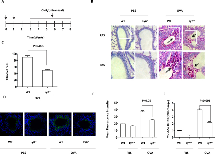
Figure 1. Mucus hypersecretion and muc5ac transcripts in OVA-induced Lyntg mice.
(A) Protocol for sensitization and challenge of Lyntg mice and WT mice with OVA (n = 8 mice for each group). (B) PAS staining of epithelial goblet cells in the lungs of WT and Lyntg mice exposed to OVA or PBS (original magnification, x200). (C) The PAS-positive cell percentage was quantified in 10 random fields (original magnification, x200). (D) Muc5ac in 16HBE cells was determined by immunofluorescence (original magnification, x200, the blue color is for DAPI). (E) The intensity of muc5ac was quantified. (F) The mRNA expression of muc5ac was determined in the lungs of Lyntg mice and WT mice using qRT-PCR (n = 3 mice for each group).

