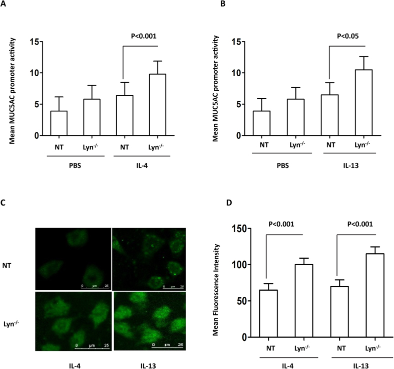
Figure 4. The levels of the MUC5AC transcript and protein in IL-4/IL-13-treated Lyn−/− 16HBE cells in vitro (Lyn−/− for siRNA treated cells, NT for untransfected cells).
MUC5AC activation was determined using a luciferase promoter assay. The human airway epithelial 16HBE cells were cotransfected with the MUC5AC promoter-luciferase plasmid and Lyn siRNA. (A) The cells were then stimulated with 1 ng/ml IL-4 for 24 hours, and the luciferase activity was measured. (B) The cells were stimulated with 1 ng/ml IL-13 for 24 hours, and the luciferase activity was measured. (C) Immunohistochemical analysis of MUC5AC in Lyn−/− cells and untransfected (NT) cells exposed to IL-4 or IL-13 for 24 hours. (D) Quantitation of the fluorescence intensity of MUC5AC per micrometer in 10 random fields as shown in (C). The results are shown as the mean ± s.d. All data are representative of three experiments, and a statistical analysis was performed.

