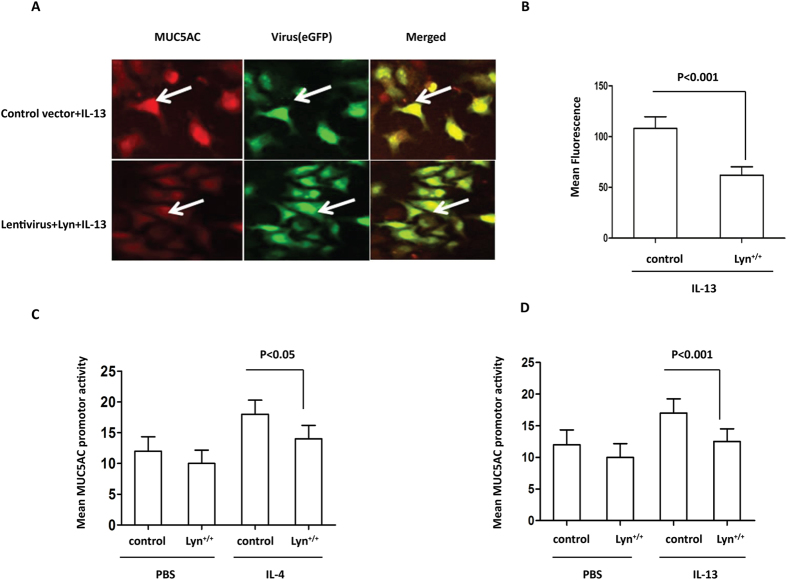
Figure 5. The levels of the MUC5AC transcript and protein in IL-4/IL-13-treated Lyn+/+.16HBE cells in vitro.
MUC5AC activation was determined using a luciferase promoter assay. We infected 16HBE cells with the Lyn-eGFP-expression lentiviral vector and control vector respectively (108 viral particles per well of a 24-well plate). (A) The cells were then stimulated with 1 ng/ml IL-13 for 24 hours. Immunohistochemical analysis of MUC5AC in Lyn+/+cells and control cells. The arrows indicate relevant markers in the positively stained cells: MUC5AC (red); eGFP or Lyn-eGFP (green). One representative experiment out of three is shown (original magnification x400). (B) Quantitation of the fluorescence intensity of MUC5AC per micrometer in 10 random fields of the images as shown in (A). (C) A MUC5AC promoter construct was transfected into control cells and Lyn+/+ cells. The cells were then stimulated with 1 ng/ml IL-4 for 24 hours. (D) The MUC5AC promoter construct was transfected into control cells and Lyn+/+ cells. The cells were then stimulated with 1 ng/ml IL-13 for 24 hours. The results are shown as the mean ± s.d. All data are representative of three experiments, and a statistical analysis was performed.

