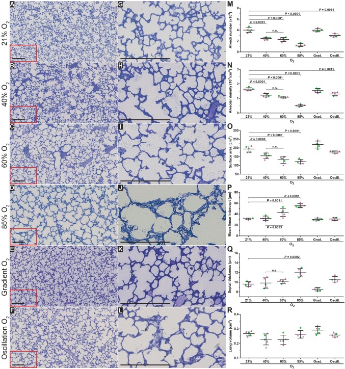
Fig. 2.
Optimising oxygen exposure levels to model bronchopulmonary dysplasia in newborn mice. Histological images were obtained from the lungs of mice subjected to the six oxygen exposure protocols illustrated in Fig. 1A. (A-F) Low-magnification images from the lungs, which were embedded in glycol methylacrylate plastic after fixation with buffered paraformaldehyde/glutaraldehyde and treatment with sodium cacodylate, osmium tetroxide, and uranyl acetate, then stained with Richardson's stain. (G-L) Higher-magnification images derived from part (demarcated by the red box) of the corresponding images to the left, to highlight changes in alveolar septal wall thickness. Each image is representative of lung sections obtained from four other mouse pups within each experimental group (n=5, per group). Scale bars: 200 µm. (M-Q) Design-based stereology was employed to assess (M) total number of alveoli in the lung, (N) alveolar density, (O) gas exchange surface area, (P) mean linear intercept, and (Q) alveolar septal wall thickness. (R) The lung volume was estimated by the Cavalieri method. In panels M-R, • denotes male animals, ▴ denotes female animals. Data represented as mean±s.d. Data comparisons were made by one-way ANOVA with Tukey's post hoc test. Significant P-values are indicated in the graphs; n.s., not significant (P≥0.05).

