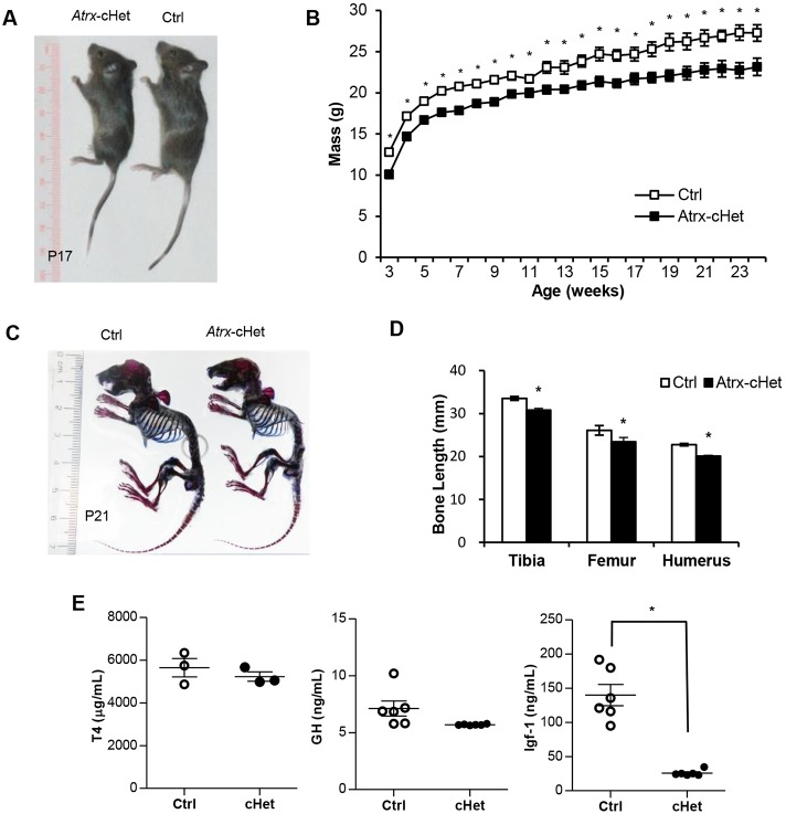
Fig. 2.
Atrx-cHet mice have reduced body weight and low circulating IGF-1. (A) Atrx-cHet female mice are smaller than littermate-matched controls at P17. (B) Growth curve of mice from 3 to 24 weeks of age (n=13; *P<0.05). Data are represented as means±s.e.m., two-way repeated-measures ANOVA with Benjamini–Hochburg post-hoc test. (C) Skeletal stains of P21 control and Atrx-cHet mice showing cartilage (blue) and bone (red). (D) The lengths of long bones of Atrx-cHet mice (n=21) are decreased compared with control mice (n=19). (E) Circulating concentrations of T4, GH and IGF-1 in Atrx-cHet and control mice (n=3) at P17. Data are represented as means±s.e.m., Student's two-tailed, unpaired t-test. *P<0.05.

