Figure 3.
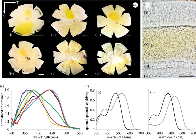
Novel intraocular filter in lanternfishes. (a) Diversity in the yellow pigmentation distribution across the retina of six species of lanternfishes: Gonichthys tenuiculus (i), Hygophum proximum (ii), S. rufinus (iii), Symbolophorus evermanni (iv), Myctophum lychnobium (v) and Myctophum obtusirostre (vi). T, Temporal; V, ventral. (b) Location and distribution of the yellow pigmentation in the retina of G. tenuiculus. PRL, Photoreceptor layer; ONL, outer nuclear layer; INL, inner nuclear layer; GCL, ganglion cell layer. Scale bar, 50 µm, (c) Normalized corrected absorbance spectra of the yellow pigment in each of the six species presented in (a). (d) Modelling of the quantal spectral sensitivity of the two visual pigments measured in M. obtusirostre, without the presence of the yellow pigmentation (i) and with the yellow pigmentation associated with the long-wave-shifted visual pigment (527 nm) (ii). Black line: visual pigment 473 nm, grey line: visual pigment 527 nm. Adapted from [47] with the permission of S. Karger AG, Basel.

