Fig. 1.
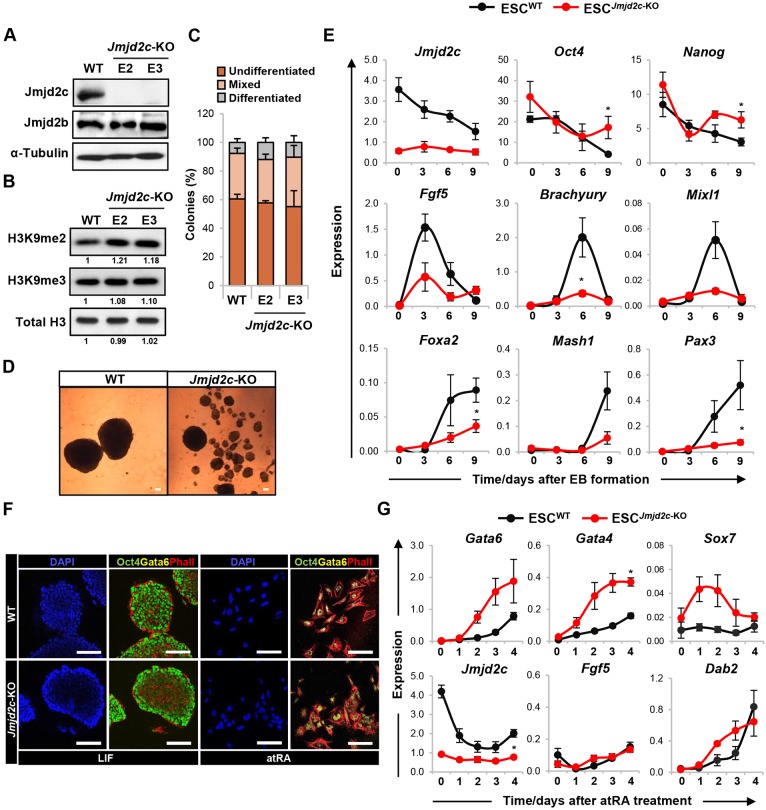
Jmjd2c-knockout ESCs can self-renew but fail to differentiate into somatic lineages. (A) Western blot using anti-Jmjd2c and anti-Jmjd2b antibodies of whole-cell extracts from wild-type (WT) JM8-ESCs and Jmjd2c-knockout (Jmjd2c-KO) cell lines (E2 and E3). α-Tubulin is used as loading control. (B) Western blots showing bulk levels of H3K9me2, H3K9me3 and total histone H3 in acid-extracted histone lysates from wild-type and Jmjd2c-KO cells. Signal quantification is presented relative to wild type. (C) Ability of wild-type and Jmjd2c-KO cells to self-renew. Cells were plated at low density and cultured for 5 days with LIF. Colonies were scored as undifferentiated, mixed or differentiated based on alkaline phosphatase activity. Data represent mean±s.e.m. of four experiments. (D) Phase-contrast images of day 9 embryoid bodies (EBs) formed from wild-type and Jmjd2c-KO (E3) ESCs. Scale bars: 100 µm. (E) Expression profiling of Jmjd2c, pluripotency-associated (Nanog, Oct4), epiblast (Fgf5), mesoderm (brachyury, Mixl1), endoderm (Foxa2) and neuroectoderm (Mash1, Pax3) markers during EB-mediated differentiation, as assessed by RT-qPCR and normalised to housekeeping genes. Data represent mean±s.e.m. of at least three experiments. *P<0.05; Mann–Whitney U-test at peak time-points. (F) Immunofluorescence staining for Oct4 (green), Gata6 (yellow) and phalloidin (red) in wild-type and Jmjd2c-KO (E3) ESCs maintained under proliferative conditions or upon 1 µM retinoic acid (atRA) addition and LIF removal for 4 days. Scale bars: 100 µm. (G) Transcript levels of Gata6, Gata4, Sox7 and Dab2 (PrE markers), Jmjd2c and Fgf5, as assessed during atRA-induced differentiation. Expression is normalised to housekeeping genes and data show mean±s.e.m. of three experiments. *P<0.05; Mann–Whitney U-test at day 4.

