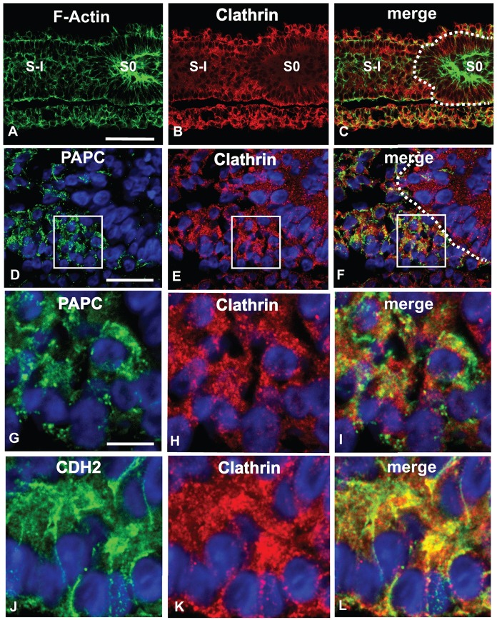
Fig. 6.
PAPC, CDH2 and clathrin colocalize in the anterior PSM. (A-C) Immunostainings of parasagittal sections of E2 chicken embryos anterior PSM stained with Phalloidin (F-actin; A, green) and an antibody against clathrin (B, red) (n=4). S-I/0, somite -I/0. Scale bar: 50 µm. (D-L) Higher resolution images comparing clathrin localization with PAPC (D-I) and CDH2 (J-L) protein immunolocalization during somite formation. G-I show higher magnifications of the boxed areas in D-F, respectively. Nuclei are counterstained (blue). Parasagittal sections, anterior to the right. Scale bars: 20 µm (D-F); 10 µm (G-L).

