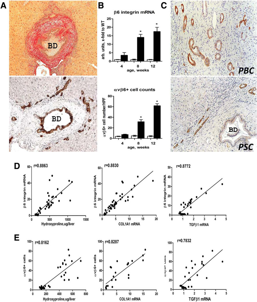
Fig. 1.
Expansion of integrin αvβ6+ ductal cells in Mdr2−/− mice parallels fibrosis progression and is present in human biliary cirrhosis. (A) Typical “onion skin” lesion around large αvβ6+ bile duct, with clusters of αvβ6+ cells resembling reactive cholangiocytes/hepatic progenitors (“ductular reaction”) at the interface of fibrotic septa and hepatic lobule. Representative images from 12-week-old Mdr2−/− mice, sirius red (upper image) and αvβ6 immunohistochemistry (lower image). (B) Increases in integrin β6 mRNA expression and αvβ6+ cell numbers parallel hepatic fibrosis progression from week 4 (incipient fibrosis) to week 12 (advanced fibrosis) in Mdr2−/− mice (closed bars) compared to Mdr2+/+ healthy littermate controls (open bars). αvβ6-positive cells were counted in 10 random portal fields of four individual mice per time point at ×200 magnification. (C) Survey of αvβ6 integrin in human explant livers with end-stage biliary cirrhosis due to PBC and PSC demonstrates a ductular cell pattern of expression similar to Mdr2−/− mice. Representative immunohistochemistry images (PSC n = 3, PBC n = 4) at ×200 original magnification. (D,E) Hepatic αvβ6+ cell counts and integrin β6 mRNA levels strongly correlate with degree of fibrosis (hydroxyproline content) and fibrosis-related gene expression (COL1A1 and TGFβ1, Spearman test r as indicated) in Mdr2−/− mice analyzed at 4, 8, and 12 weeks of age. Abbreviations: BD, bile duct; HPF, high-power field; WT, wild type.

