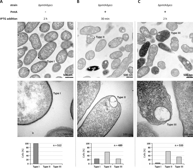
FIG 5 .
Intracellular structures are formed upon overproduction of PmtA in A. tumefaciens. (A to C) A. tumefaciens ΔpmtA Δpcs cells carrying pTRC200 empty vector (A) or pTRC200_pmtA expression plasmid (B and C) were grown to the logarithmic growth phase (OD600 of 1.5). Protein production was induced using 100 µM IPTG. Cell morphology was analyzed by transmission electron microscopy 30 min and 2 h after the induction of PmtA overproduction. Bacterial cells were fixed, embedded, ultrathin sectioned, and analyzed using a Philips CM100 electron microscope. Images were taken with a CCD camera (Orius SC600; Gatan, Inc.). Three different cell types were observed. The bottom images show magnifications of selected cells, and the graphs below show quantification of the cell types (n is the total number of counted cells). Type I, initial morphology of A. tumefaciens ΔpmtA Δpcs cells; type II, cells carrying vesicle-like structures; type III, cells with vesicle-like structures, high electron density, and damaged cell membrane.

