Fig. 2.

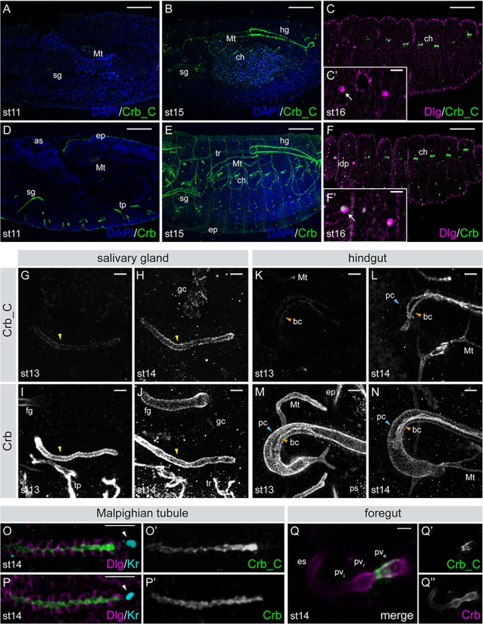
Expression of Crb isoforms during embryogenesis. (A-F) Lateral view of wild-type embryos at stage 11 (A,D), stage 15 (B,E), and stage 16 (C,F) stained with α-Crb_C or α-Crb (green) and α-Discs large (Dlg; magenta); nuclei (DAPI, blue). Scale bars: 50 µm. (C′,F′) Higher magnification of the precursors of the imaginal disc (idp; arrow). Scale bars:10 µm. (G-N) Dorsal views of stage 13 and 14 embryos showing the salivary gland (sg; G-J, yellow arrowheads) and hindgut (hg; K-N). Crb_C expression increases during sg development. In the hg, expression of Crb_C is first detected in the boundary cells at stage 13 (orange arrowheads in K-N). Crb_C expression level gradually increases during embryogenesis (L). The principal cells (pc; blue arrowheads) express very low levels of Crb_C (L). (O,P) Dorsal view of the Malpighian tubules (Mt) at stage 14 stained with α-Crb_C (O,O′) or α-Crb (P,P′). Dlg (magenta) marks the lateral membranes, Krüppel (Kr, cyan) the nucleus of the distal tip cell (white arrowheads). Crb_C is predominantly expressed at the distal tip. (Q) Ventral view of the foregut stained with α-Crb_C (green, Q′) and α-Crb (magenta, Q″). α-Crb_C only stains the external portion of the proventriculus (pve), while α-Crb stains all parts. as, amnioserosa; bc, boundary cells; ch, chordotonal organs; ep, epidermis; es, esophagus; fg, foregut; gc, garland cells; hg, hindgut; idp, imaginal disc precursors; Mt, Malpighian tubule; pc, principal cells; ps, posterior spiracle; pve, external portion of the proventriculus; pvi, internal portion of the proventriculus; pvr, recurrent portion of the proventriculus; sg, salivary gland; tp, tracheal pits; tr, tracheal tree. Anterior to the left. Scale bars: 10 µm.
