Fig. 7.

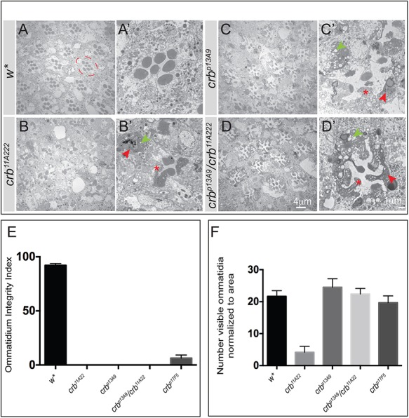
crb_C mutant photoreceptor cells undergo light-dependent degeneration. (A-D′) Representative TEM images of retinal cross sections of adult flies of the indicated genotypes after 7 days continuous light exposure. A-D are overview images, A′-D′ are higher magnifications of one ommatidium. The stereotypic trapezoid arrangement of the seven rhabdomeres (w*) is lost in all crb alleles. Signs of degeneration include loss of rhabdomeric integrity (red asterisk), accumulation of electron dense debris (red arrowheads) and extensive vacuolization (green arrowheads). The dashed red circle in A outlines one ommatidium. Scale bars: 4 µm (A,B,C,D), 1 µm (A′,B′,C′,D′). (E) Bar graph depicting the mean Ommatidium Integrity Index (OII)±s.e.m. Ommatidial integrity index is the percent ratio of ommatidia (with no obvious signs of intracellular debris and vacuolization) to the total number of ommatidia normalized to area for the genotypes indicated on the x-axis. The significantly reduced OII in all crb alleles highlights retinal degeneration in these genotypes. (F) Bar graph depicting the mean number±s.e.m. of remnant ommatidia, normalized to area, in each of the genotypes indicated on the x -axis. Although there is degeneration in all crb mutant alleles, the phenotype is most severe in the loss-of-function allele crb11A22 (large clone mosaics), evident from the reduced number of identifiable ommatidia per unit area (compare with sections shown in the TEM panel above). Sample size consists of three biological replicates, from which at least 100 ommatidia were evaluated (with the exception of crb11A22, in which most of the ommatidia completely degenerate and only 25 ommatidia could be identified for quantification).
