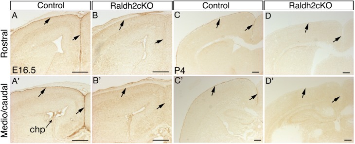
Fig. 1.
Tamoxifen-induced ablation of Raldh2 in the developing meninges. (A,A′,C,C′) Immunodetection of RALDH2 on coronal sections of the brain of a control (Raldh2flox/flox;CMV-βactin-Cre-ERT20) mouse at E16.5 (A,A′) and at P4 (C,C′). Sections are shown at a rostral (A-D) and more caudal (A′-D′) level of the brain, showing RALDH2 expression in the meningeal layer overlying the cerebral cortex (arrows), and in choroid plexus (chp). (B,B′,D,D′) Comparative views of the brain of a Raldh2cKO (Raldh2flox/flox; CMV-βactin-Cre-ERT2+) mutant, showing absence of RALDH2 signal. Tamoxifen was administered at E10.5. Scale bars: 250 μm.

