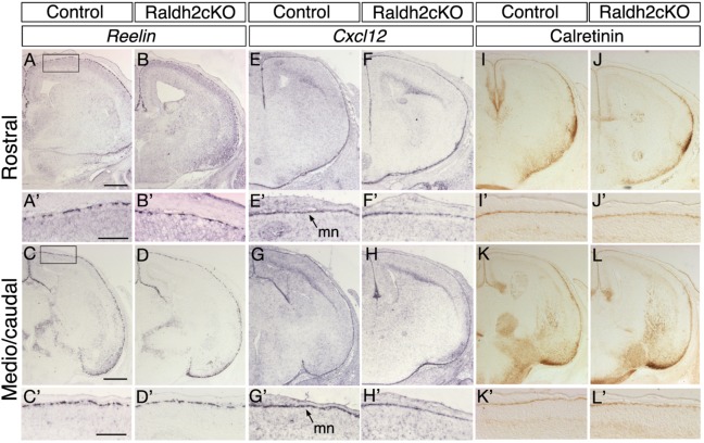
Fig. 2.
Analysis of Cajal–Retzius cells and developing meninges in Raldh2cKO mice. Comparative, coronal E16.5 brain sections are shown at two levels, rostral (upper panels) and more caudal (lower panels). In situ hybridisation for Reelin (A-D′) and immunolabellings for Calretinin (I-L′) show that their distribution in Cajal–Retzius cells of the cortical marginal zone of control embryos is unchanged in Raldh2cKO embryos. In situ hybridisation for Cxcl12 (E-H′), a marker of the developing meninges, also shows a normal distribution in Raldh2cKO embryos. mn, meninges. Scale bars: 500 μm (A-L), 250 μm (A′-L′).

