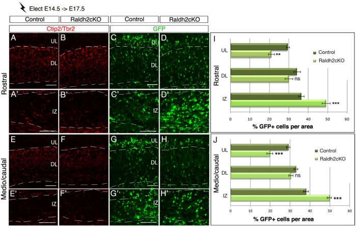
Fig. 8.
Lack of RA perturbs cell migration of late-born cortical neurons. Brain sections from E17.5 embryos electroporated at E14.5 with the GFP reporter were analysed by immunolabelling for Tbr2 and Ctip2 (A-B′,E-F′) and for GFP-expressing cells (C-D′,G-H′), at rostral (A-D′) and caudal levels (E-H′) of the cortex. (I,J) Histograms show the percentage of GFP-positive cells in control and Raldh2cKO animals per upper layer (UL), deeper layer (DL) and intermediate zone (IZ) of the developing cortex. Data presented as mean±s.e.m.; n=5 brains; **P<0.01, ***P<0.001; ns, not significant by two-way ANOVA. Scale bars: 100 μm.

