
Figure 1. Analysis of miR-101 expression in RCC clinical specimens and functional analysis of miR-101 transfection in 786-O and Caki-1 cells.
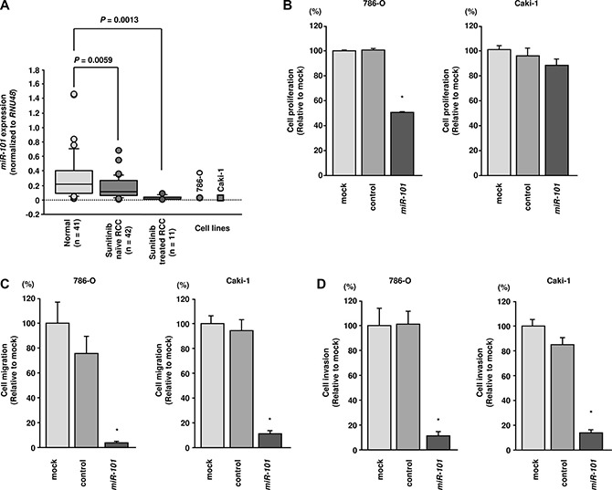
(A) Expression levels of miR-101 in RCC clinical specimens. RNU48 was used for normalization. (B) Cell proliferation was assessed 72 h after transfection with miR-101 using XTT assays. (C) Cell migration was assessed 48 h after transfection with miR-101 using uncoated Transwell polycarbonate membrane filters. (D) Cell invasion was assessed 48 h after transfection with miR- 101 using Matrigel invasion assays. *P< 0.0001. The bars indicate SDs.