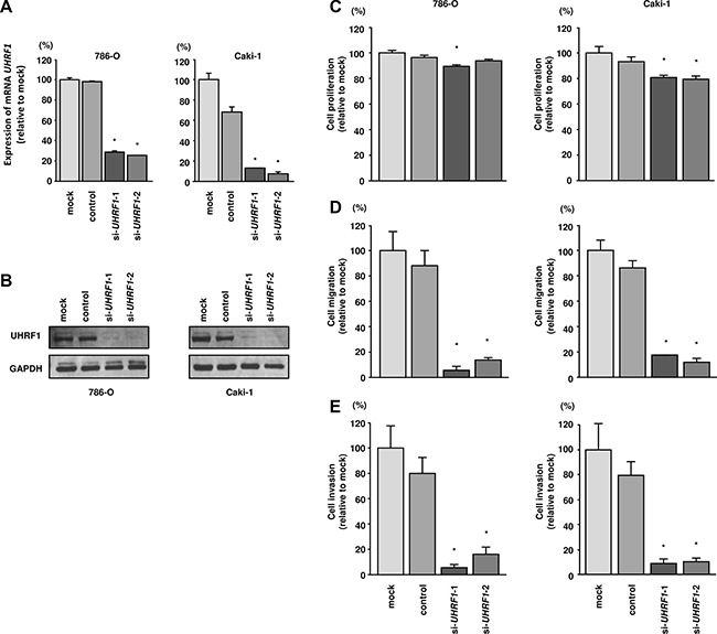
Figure 5. Effects of UHRF1 knockdown in RCC cells and impact of UHRF1 expression on clinical RCC specimens.

(A) UHRF1 mRNA expression was determined at 72 h after transfection with si-UHRF1. GUSB was used as an internal control. (B) UHRF1 protein expression was evaluated by western blotting at 72 h after transfection with si-UHRF1. GAPDH was used as a loading control. *P < 0.0001. The bars indicate SDs. (C) Cell proliferation was assessed 72 h after transfection with si-UHRF1 using XTT assays. (D) Cell migration was assessed 48 h after transfection with si-UHRF1 using uncoated Transwell polycarbonate membrane filters. (E) Cell invasion was assessed 48 h after transfection with si-UHRF1 using Matrigel invasion assays. *P < 0.0001. The bars indicate SDs.
