
Figure 4. XOR suppression inhibits genotoxic stress-induced uric acid production and NKG2D ligand expression.
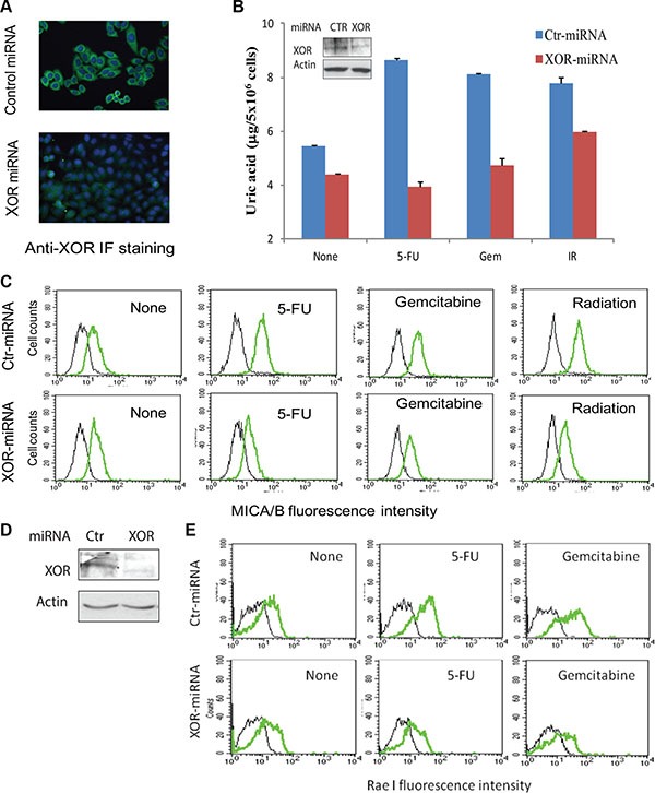
(A) Immunofluorescence (IF) staining of XOR. HeLa cells stably transfected with control miRNA (Ctr-miRNA) or XOR-miRNA were analyzed for XOR expression by IF staining with an anti-XOR antibody as described in Figure 1A. Nuclei was stained with DAPI. (B and C) XOR knockdown blocks uric acid production and MICA/B expression. Ctr-miRNA and XOR-miRNA-transfected cells were exposed to 5-FU (10 μM), gemcitabine (2 μM), or radiation (40 Gy). Cells were harvested at 24 hr and analyzed for intracellular uric acid levels using a uric acid assay kit (B) or for MICA/B expression by FACS (C). Black line, isotype control; Green line, MICA/B. Inset in (B): XOR expression analyzed by Western blot. Actin was included as a loading control. (D) Western blot analysis of XOR expression in Ctr-miRNA- and XOR-miRNA-transfected RCAS-Neu cells. (E) XOR knockdown blocks genotoxic stress-induced Rae I expression. Ctr-miRNA- and XOR-miRNA-transfected RCAS-Neucells were exposed to 5-FU (10 μM) or gemcitabine (2 μM) and analyzed for Rae I expression 16 hr later by FACS analysis.