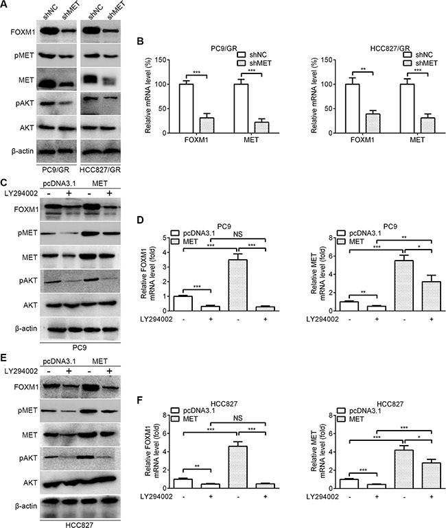
Figure 5. FOXM1 is activated by MET/AKT signaling pathway.

(A) Western blot analysis for FOXM1, pMET, MET, pAKT and AKT in PC9/GR and HCC827/GR that were transfected with MET shRNA. (B) Quantitative real-time PCR analysis for FOXM1 and MET in PC9/GR and HCC827/GR that were transfected with MET shRNA. (C–F) PC9-MET and HCC827-MET cells were treated with LY294002 for 6 hours, the protein levels of FOXM1, pMET, MET, pAKT and AKT were analyzed by western blot analysis, and the mRNA levels of FOXM1 and MET were analyzed by quantitative real-time PCR analysis. Each bar represents the mean ± SD. P values were calculated using Student's t-test (*P < 0.05, **P < 0.01, ***P < 0.001, NS, nonsignificant).
