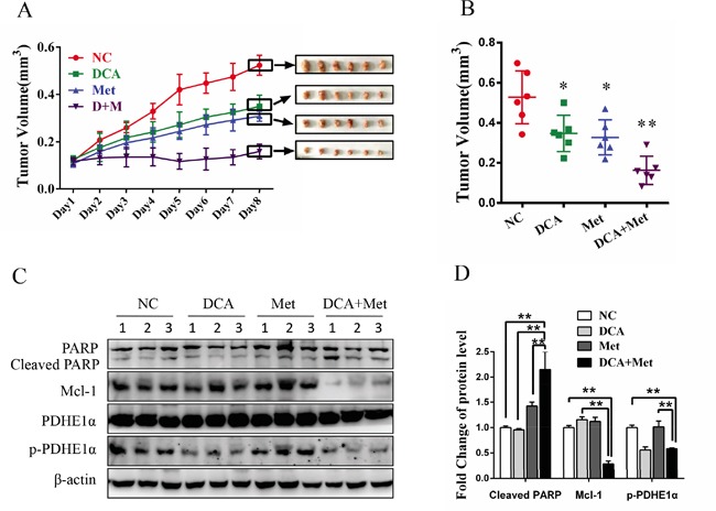
Figure 6. DCA and Met collaboratively repress the growth of ovarian cancer cells in vivo.

A-D. 5×10⁶ SKOV3 cells in 150 μL PBS were implanted into the right axillae of each nude mouse. When palpable tumors were formed, the mice were randomized into 4 groups (n = 6 per group). Then the mice were intraperitoneally injected everyday with DCA (50 mg/kg/d) plus Met (100 mg/kg/d) or each alone for 8 days, taking PBS as control. The xenograft tumor size was monitored every day (volume = width2×length×1/2) (A). After excised from the mice, the xenograft tumors were photographed (A) and their volumes were showed in (B). The levels of cleaved PARP, Mcl-1, total PDHE1α and p-PDHE1α were measured by Western blot (C), and the ratios of the corresponding proteins to β-actin were calculated (D). *,P<0.05; **,P<0.01.
