
Figure 2. Gy1 can specifically bind and internalize into PSMA positive cancer cells.
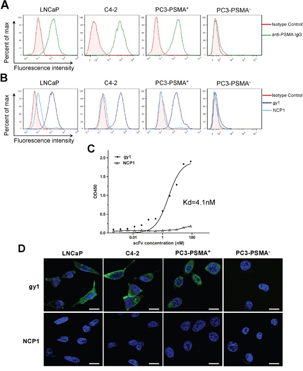
A. Flow cytometry analysis to show the PSMA expression on different prostate cancer cells. B. Flow cytometry analysis to show the binding of gy1 to PSMA positive cancer cells. LNCaP, C4-2, PC3-PSMA+and PC3-PSMA− cells were incubated with 100 nM of gy1 and followed by FITC-conjugated secondary antibody. NCP1 was used as negative control. C. Cellular ELISA to show the binding affinity of gy1. The Kd was calculated using non-linear regression analysis of a one-site binding hyperbola equation of GraphPad Prism 5.0 software. Representative result was shown from 3 independent experiments. D. Immunofluorescence staining to show the internalization of gy1 into PSMA positive cancer cells. Gy1 was incubated with LNCaP, C4-2, PC3-PSMA+ and PC3-PSMA− cells for 2 h before immunofluorescence staining. Scale bar = 25 μm.