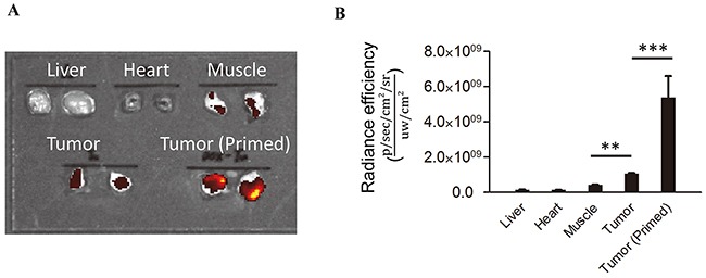
Figure 4. exDNA is enriched in tumors.

The relative amounts of exDNA in normal tissues and 4T1 tumors with and without treatment with 3E10EN/DOX-NPs was determined by Picogreen staining. The amount of exDNA in untreated tumors was 7.5, 11.7 and 2.5 times greater than what is found in the liver, heart, and muscle. Treatment of the mice with 3E10EN/DOX-NPs increased the amount of exDNA in tumors by 5.1 fold compared to tumors in untreated mice. A representative image of exDNA in indicated tissues is shown in A. and a quantitative analysis of exDNA present in the indicated tissues (n=5) is shown in B. The data are presented as mean +/− SD. **: P < 0.01. ***: P < 0.001.
