
Figure 1. Disruption of the FA pathway induces a short-lived response to oncogenic stress in vitro.
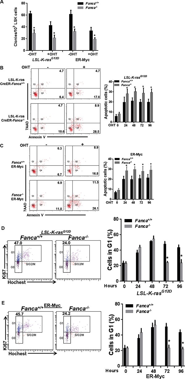
(A) Oncogenic stress compromises colony formation capacity of FA HSPCs. LSK cells (Lin−Sca1+c-kit+ cells) isolated from LSL-Fanca+/+/K-ras/CreER and LSL-Fanca−/−/K-ras/CreER mice, or retroviral vector MSCV-IRES-MycER transduced LSK cells from Fanca+/+ or Fanca−/− mice were in vitro culture in the presence of 4-OHT for 48 hours followed by plating in cytokine-supplemented methycellulose medium. Colonies were enumerated on day 7 after plating. Results are means ± standard deviation (SD) of 3 independent experiments (n = 9 per group). (B) K-ras activation induces apoptosis in FA HSCs. LSK cells (Lin−Sca1+c-kit+ cells) isolated from LSL-Fanca+/+/K-ras/CreER and LSL-Fanca−/−/K-ras/CreER mice were subjected to Flow cytometric analysis for apoptosis by Annexin V/7AAD staining at different time points. Representative images at time 0 and 24 h after 4-OHT induction (left) and quantification (right) were shown. Results are means ± standard deviation (SD) of 3 independent experiments (n = 6 per group). (C) Myc activation induces apoptosis in FA HSCs. Retroviral vector MSCV-IRES-MycER transduced LSK cells from Fanca+/+ or Fanca−/− mice were subjected to Flow cytometric analysis for apoptosis by Annexin V/7AAD staining at different time points. Representative images at time 0 and 24 h after 4-OHT induction (left) and quantification (right) were shown. Results are means ± standard deviation (SD) of 3 independent experiments (n = 9 per group). (D) Activation of K-ras leads to short-lived G1 arrest in FA cells. Cells described in (B) were cultured in the presence of 4-OHT for 2 hours then released in fresh medium for the indicated time intervals, followed by cell cycle profiling by Hochest33324/Ki67 staining. Representative images (left) and quantification (right) were shown. Results are means ± standard deviation (SD) of 3 independent experiments (n = 6 per group). (E) Activation of Myc leads to short-lived G1 arrest in FA cells. Cells described in (C) were cultured in the presence of 4-OHT for 2 hours then released in fresh medium for the indicated time intervals, followed by cell cycle profiling by Hochest33324/Ki67 staining. Representative images (left) and quantification (right) were shown. Results are means ± standard deviation (SD) of 3 independent experiments (n = 9 per group).