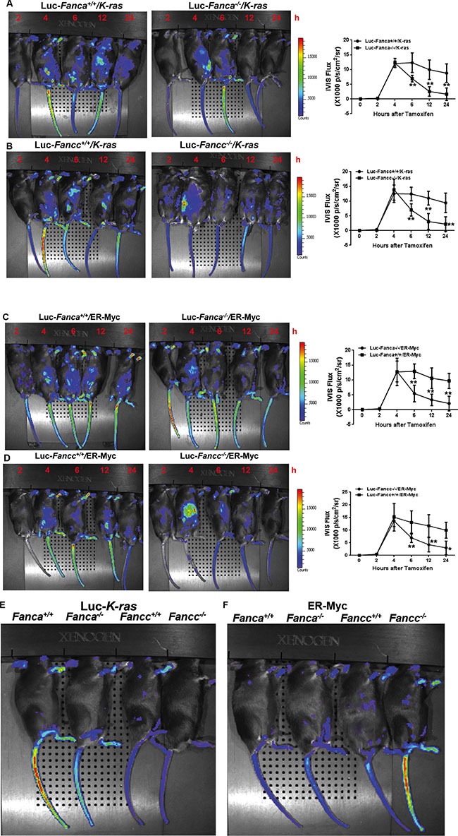
Figure 2. Disruption of the FA pathway induces a short-lived response to oncogenic stress in vivo.

(A, B) FA mice exhibit short-lived response to K-ras activation. 1,000 LSK cells from Luc-LSL-K-ras/CreER-Fanca+/+ or 2,000 LSK cell from Luc-LSL-K-ras/CreER-Fanca−/− mice (A) along with 3 × 10⁵ BM cells from congenic BoyJ mice were transplanted into lethally irradiated BoyJ recipients. 4-month post BMT, the recipients were i.p injected with single dose of Tamoxifen followed by IVIS imaging at the indicated time points. Luminescence scale is in p/s/cm2/sr. Similar experiments were conducted on Fancc−/− mice (B). (C, D) FA mice exhibit short-lived response to MycER activation. 1,500 retroviral vector MSCV-IRES-MycER transduced Luc-Fanca+/+ cells or 3,000 transduced Luc-Fanca−/− cells (C) along with 3 × 10⁵ BM cells from congenic BoyJ mice from recipient mice were transplanted into lethally irradiated BoyJ recipients. 4-month post BMT, the recipients were i.p injected with single dose of Tamoxifen followed by IVIS imaging at the indicated time points. Luminescence scale is in p/s/cm2/sr. Similar experiments were conducted on Fancc−/− mice (D). The bioluminescent image signals were quantified using LiveImage Pro. 2.0 software. Results are means ± standard deviation (SD) of 3 independent experiments (n = 6 per group). (E, F) Minimum bioluminescence of Luc mice without tamoxifen injection (the “0” time-point controls). Luminescence scale is in p/s/cm2/sr.
