
Figure 4. Restoration of ERα expression renders endometrial cancer cells susceptible to megestrol acetate treatment.
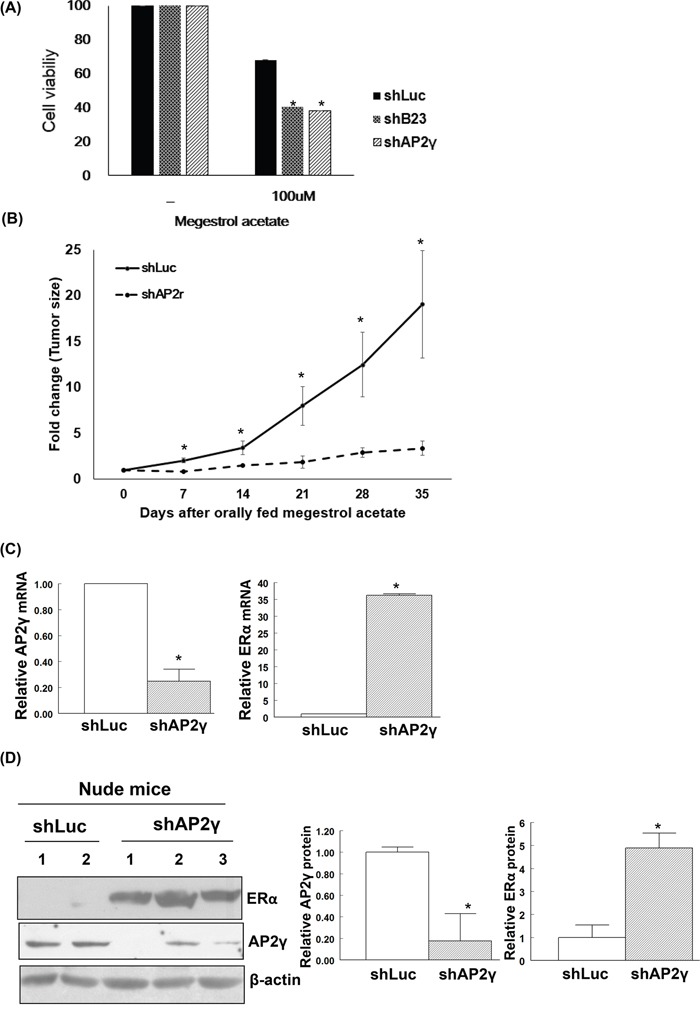
A. Human endometrial cancer Ishikawa/shLuc (shLuc), Ishikawa/shB23 (shB23), and Ishikawa/shAP2γ (shAP2γ) cells were treated with 100 nM megestrol acetate for 72 h and assayed by MTT. Nude mice experiments are shown in (B-D). B. Knock-down cell lines Ishikawa/shLuc (shLuc), Ishikawa/shB23 (shB23), and Ishikawa/shAP2γ (shAP2γ) were inoculated into nude mice; tumor volumes were subsequently measured on a weekly basis. No tumor formation was observed when animals were inoculated with the stable B23 knock-down cell line. Tumor-bearing nude mice orally received megestrol acetate (10 mg/kg) dissolved in cooking oil (5 days per week). The points depict the means (± standard deviations) of tumor volumes for megestrol acetate-treated (n = 3) and control animals (n = 2). *P < 0.05 compared with controls. After treatment with megestrol acetate, tumors derived from Ishikawa/shLuc (shLuc) and Ishikawa/shAP2γ (shAP2γ) cells were analyzed for mRNA (C) and protein levels (D). C. RNAs were analyzed with real-time qPCR with the reported primers. In the quantitative bar graph, the results are expressed as means ± standard errors of the mean. D. Cell lysates immunoblotted with ERα and AP2γ antibodies. The presence of an equal amount of proteins in each lane was confirmed with β-actin. In the quantitative bar graph, the results are expressed as mean ± standard error of the mean.