
Figure 8. PRIMA-1MET decreased relative cell number of GSCs irrespective of p53 status.
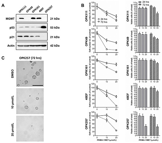
A. Western blotting analysis showing expression of MGMT, p53 and p21 in OPK111, OPK49, OPK161, 48EF and OPK257 GSCs. Actin was used as a loading control. B. Analysis of the cytotoxic effect of PRIMA-1MET (10 or 20 μM) on OPK111, OPK49, OPK161, 48EF and OPK257 GSCs using trypan blue exclusion assay and automated cell counting to determine the percentage of relative number of cells in PRIMA-1MET-treated conditions relative to DMSO control at each time point (24 or 72 hours following initiation of a 24-hour treatment with PRIMA-1MET) (left) and the ratio of viable cells (% relative to total cell number in each experimental condition) (right) in the indicated cell lines. Data on graphs represent the mean values ± SD. C. Representative micrographs of OPK257 GSCs (original magnification 200X) treated with PRIMA-1MET (10 or 20 μM) or DMSO control at 72-hour time point. Scale bar = 200 μm.