
Figure 1. OSM induced the tyrosine phosphorylation and nuclear translocation of STAT1 and STAT3.
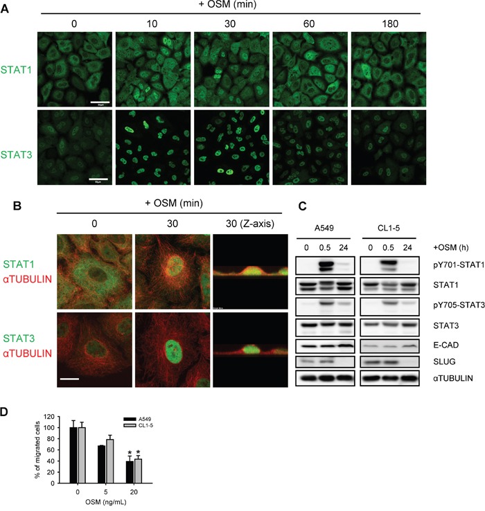
A, B. A549 cells treated with 20 ng/mL OSM for the indicated durations were subjected to immunofluorescent staining of STAT1, STAT3 (green) and αTUBULIN (red), and observed by confocal microscope. Scale bar = 50 μm in (A), 10 μm in (B). C. A549 and CL1-5 cells were subjected to Western blotting analysis after 0.5 and 24 hours of incubation in 20 ng/mL OSM. pY701-STAT1 and pY705-STAT3 indicated the phosphorylated STAT1 and STAT3, respectively. D. A549 and CL1-5 cells were subjected to the migration assay in the presence of 5 or 20 ng/mL OSM. Sterile water was used as control vehicle for non-treated cells. The results are presented as the percentage of migrated cells treated with OSM relative to non-treated control.