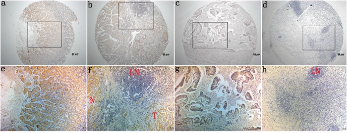
Figure 2. Immunohistochemical staining for Barx2 expression in normal gastric mucosa and cancerous tissues.

Barx2 protein expression was significantly lower in gastric cancer tissues compared with adjacent normal mucosa, with Barx2 staining observed both in the cytoplasm and nuclei of gastric cancer cells. a, e: Strong Barx2 staining in normal gastric epithelium; b, f: Intense Barx2 staining in well-differentiated gastric cancer; c, g: Weak Barx2 staining in moderately differentiated gastric cancer; d, h: Negative Barx2 staining in poorly differentiated gastric cancer; N: Strong Barx2 staining in normal gastric epithelium; T: Intense Barx2 staining in well-differentiated gastric cancer; LN: Negative Barx2 staining in lymph node; a–d. Original magnification: 50×; e–h. Original magnification: 200×.
