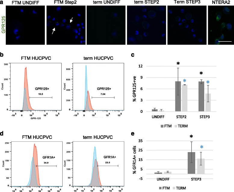
Fig. 4.

Human spermatogonial stem cell-associated markers are upregulated in first trimester (FTM) and term human umbilical cord perivascular cells (HUCPVCs) in the early steps of differentiation. a Representative micrograph of undifferentiated (UNDIFF) FTM HUCPVCs (left) and at Step 2 (second panel), undifferentiated term HUCPVCs (third panel) and at Step 2 and Step 3 (fourth and fifth panel, respectively) immunostained for GPR125 (green, undetected in undifferentiated PVCs) and counterstained with Hoechst to show live nuclei. GPR125 (green) was detected in FTM HUCPVCs and term HUCPVCs at the end of Steps 2 and 3. Human testicular cells were used as a positive control. Scale bar = 50 μm. b Representative flow cytometry analysis plots for GPR125 in FTM HUCPVCs (left) and term HUCPVCs (right), comparing the undifferentiated stage to Step 3. c Quantification of the percentage of FTM HUCPVCs and term HUCPVCs that upregulated GPR125 at Steps 2 and 3, in comparison to undifferentiated controls (p < 0.05 for all comparisons to undifferentiated). n = 3 independent lines of each FTM and term HUCPVC. d Representative flow cytometry plots for GFR1α expression analysis (also known as GDNFR) in FTM HUCPVCs (left) and term HUCPVCs (right), comparing the undifferentiated stage to Step 3. e Quantification of the percentage of FTM HUCPVCs and term HUCPVCs that upregulated GFR1α at Step 3 in comparison to undifferentiated controls (p < 0.05 for all comparisons to undifferentiated). n = 3 independent lines of each FTM and term HUCPVC
