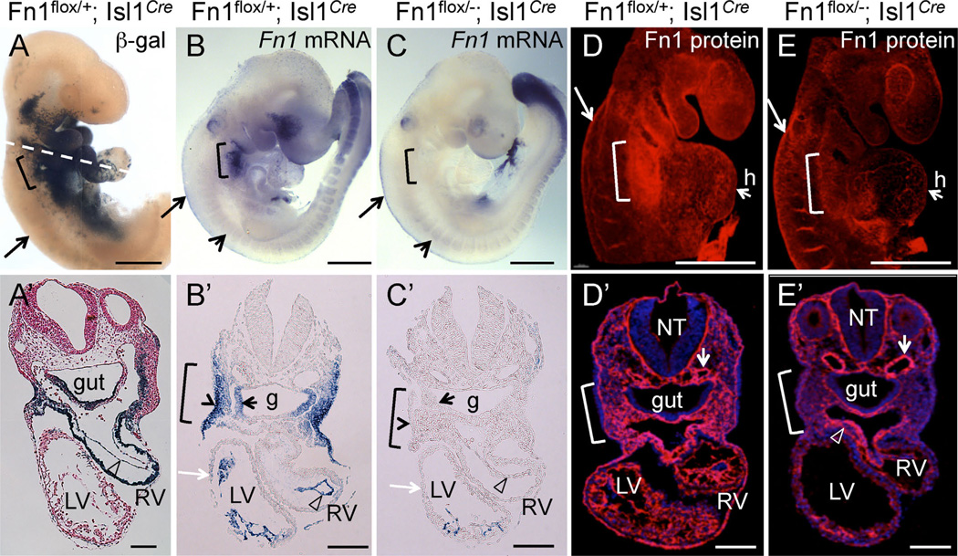
Fig. 1.
Conditional ablation of Fn1 in pharyngeal tissues using the Isl1Cre knock-in strain. All embryos are at E9.5. Brackets in all panels mark the pharyngeal region where arches 3, 4, and 6 will develop. Long arrows in (A–E) point to the neural crest. (A–A′) Fate map of Isl1+ cells and their descendants; Dashed line in A marks approximate planes of sections shown in (A′–E′), open arrowhead points to endocardium in A′–C′. (B-B′) Fn1 mRNA expression is enriched in the surface ectoderm (arrowhead in B′), pharyngeal pocket endoderm (small black arrow), mesoderm, and endocardium (arrowhead). (C-C′). Fn1 mRNA is ablated in the pharyngeal tissues following Cre-mediated recombination in Fn1flox/−; Isl1Cre mutants. Myocardium (white arrow) does not express Fn1. Short arrows in (B–C) point to somites, where Isl1Cre is not expressed and Fn1 expression is unaltered. (D) Expression of Fn1 protein is enriched in the posterior pharyngeal arches (bracket). E. Fn1 protein is depleted from the pharyngeal region in Fn1flox/−; Isl1Cre mutants. (D′–E′) Transverse sections through (D–E). Fn1 is depleted in pharyngeal arches and the heart (h) in Fn1flox−; Isl1Cre mutants (E′). Fn1 remains expressed by the neural crest-derived cells (arrowhead in E′) and by more dorsal regions, including the dorsal aorta (short arrows in D′–E′); g - gut, LV, RV - left and right ventricles, NT - neural tube. Scale bars in (A–E) are 500 µm; in (A′–E′) 100 µm.

