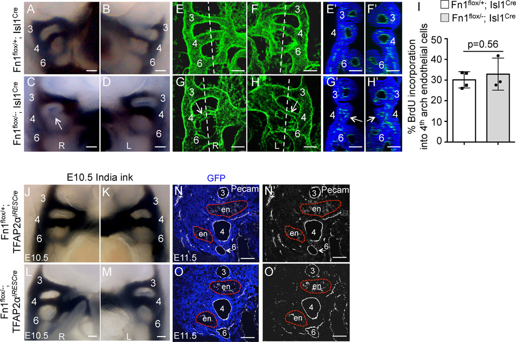
Fig. 6.
Expression of Fn1 in the Isl1-derived tissues regulates formation of the 4th PAAs. All images in (A–M) are from E10.5 embryos. PAAs are numbered. (A–D) Intracardiac India ink injections label the PAAs. All PAAs are visible in controls (A–B). The right 4th PAA (arrow) is non-patent in the Fn1flox/−; Isl1Cre mutant (C–D). E-H. Whole mount Pecam1 staining (green) and 3D–confocal image reconstruction show well-formed PAAs in controls (E–F), and bi-laterally thin 4th PAAs in the Fn1flox/−; Isl1Cre mutant, arrows (G–H). Dashed lines in (E–H) mark optical planes of section shown in (E′–H′). Pecam1 (blood vessels, green) and DRAQ5 (nuclei, blue). Arrows point to pharyngeal arches with thin/disorganized PAAs. Scale bars are 100 µm; R - right, L - left. I. BrdU incorporation into endothelial cells in controls (white bars) and mutant (gray bars) embryos; Each dot represents measurements in one pharyngeal arch; p was determined using 2-tailed, unpaired Student′s t test. (J–O′) Expression of Fn1 in the neural crest and the surface ectoderm is not required for PAA formation. (J–M) India ink injections at E10.5. All PAAs are visible in control Fn1flox/+; TFAP2αIRESCre and mutant Fn1flox/−; TFAP2αIRESCre embryos at E10.5. (N–O′) Coronal sections through E11.5 embryos show that all PAAs (numbered 3–6) are formed normally in control Fn1flox/+; TFAP2αIRESCre and mutant Fn1flox/−; TFAP2αIRESCre embryos. Neural crest is stained using anti-GFP antibodies (blue), due to the presence of the ROSAmTmG allele and endothelium is labeled with anti-Pecam1 antibodies (white). Red dashed lines outline the endoderm (en). Native GFP and tdTomato fluorescence was extinguished by boiling sections in 10 mM Citric Acid pH 6.0 as described in Methods.

