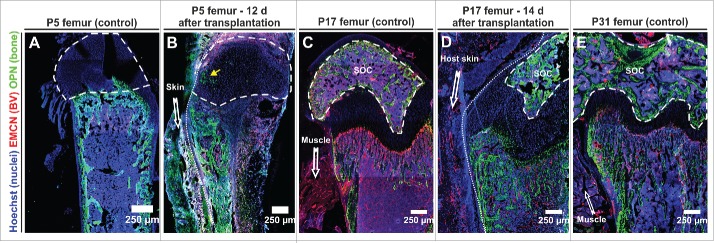
Figure 2.
The developmental fate of transplanted femurs strongly depends on the age of the graft at the time of transplantation. (A, C, E) Representative control immunofluorescence staining of neonatal femurs at the indicated age (P5, P17, P31) – 3 femurs per age category. (B) 12 days after transplantation of a P5 femur the bone marrow cavity was largely filled with new woven bone secreted by OPN+ cells. OPN+ cells were also seen to invade the epiphysis (yellow arrow). Representative figures obtained from 2 transplants. (D) Femurs collected from P17 pups maintained their open BM cavity for 14 days after transplantation and reached a developmental stage that was reminiscent of a P31 control femur. Representative figures from 4 transplants. (E). Compared to the age matched control (E), development of the secondary ossification center (SOC) in the transplanted femur was delayed. All panels depict 8 μm cryosections that were analyzed by confocal microscopy (tile-scan). Dashed line encircles the SOC, dotted line demarks border transplant host, EMCN endomucin, BV blood vessel, OPN osteopontin.

