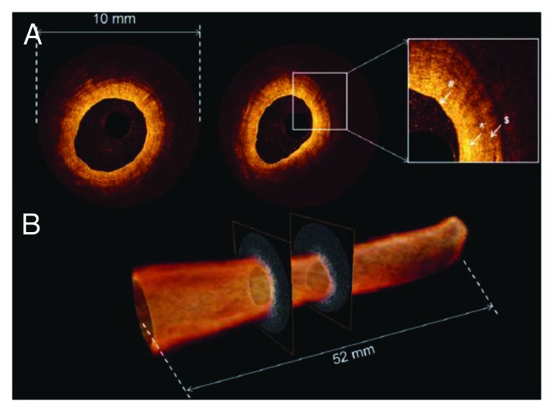
Figure 5. OCT of healthy ureter. (A) Individual OCT images obtained from volumetric OCT data set. Inset, higher magnification reveals normal ureter urothelium (pond sign), lamina propria (asterisk), and muscularis (dollar sign). (B) 520-frame volumetric data set across 52 mm trajectory along probe in approximately 5.2 s, resulting in 52 mm long by 10 mm diameter total scanned cylindrical volume. Figures and captions are adapted from reference 119 with permission.
