
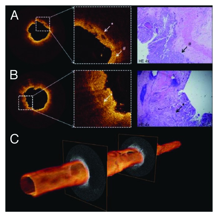
Figure 6. (A and B) Cross-sectional OCT images of proximal ureter show interruption (white asterisk) of thin dark line (white pound sign) suggesting invasive tumor. Distinction among anatomical layers was not possible. Corresponding histology reveled T3G3 urothelial carcinoma (black arrow). (C) 3D pullback of OCT built from 520 individual cross-sectional images over 5.2 cm length. Figures and captions are adapted from reference 119 with permission.