
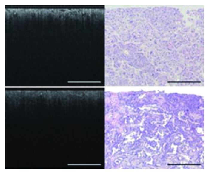
Figure 7. OCT image and corresponding light microscopy of renal carcinoma, chromophobe subtype (top panel) and papillary subtype, grade 4 (bottom panel). In the chromophobe subtype (top panel), collections of large polygonal cells arranged in trabeculae are seen as areas of intermediate brightness with intervening dark spaces on OCT. In the papillary subtype (bottom panel), elements of cuboidal cells surrounding a fibrovascular stalk were seen on light microscopy but not visible on the OCT image. Bars are 500 μm. Figures and captions are adapted from reference 122 with permission.